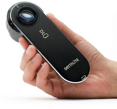
Dermoscopic examination

Dermoscopy (Dermatoscopy) Dermatology is considered by many to be “superficial medicine” because the manifestations of the disease are expressions that appear on the skin = on the surface (as opposed to other areas of medicine where the manifestations of the disease are mainly in the internal, deep organs). This uniqueness of dermatology allows dermatologists to reach a diagnosis in a very high percentage of cases based solely on observation and examination of the skin’s appearance, its lesions, and their properties, without the need for additional diagnostic tests. Traditional dermatology has therefore been for generations the art of observation and identification of details available to the eye. Therefore, it is no wonder that at the beginning of the last century, attempts began to improve the identification of details that can be extracted from observing the skin – by using devices technically designed to improve vision – by magnifying and illuminating the skin. This action of examining the skin using microscope-like devices was nicknamed “dermoscopy.” These attempts initially yielded cumbersome devices that, more than assisting in examining the skin, burdened the doctors and therefore did not gain recognition and widespread distribution. The growing recognition of the importance of early diagnosis of skin cancer lesions – and especially the melanoma epidemic, whose late diagnosis in most cases meant a death sentence – provided special motivation for the development of technical means to assist in the earliest possible diagnosis of the tumors. As a result, about 25 years ago, there was a change in dermatologists’ approach to the subject, which dramatically accelerated with technological advancements that allowed the emergence of modern dermatoscopes. These devices are portable and compact, allowing a deeper examination of skin lesions and revealing to the doctor’s eye details that were previously not visible. As a result, melanomas can now be diagnosed with an accuracy rate of over 90% through early diagnosis using dermoscopy, compared to a previous accuracy rate of only about 60-70%. An additional benefit that was initially secondary to this development was the discovery that the dermatoscope allows for more accurate diagnosis and early detection of a variety of other skin lesions and diseases, adding a significant dimension to skin examination in many additional areas. The dermatoscope has become an essential tool for the modern dermatologist, similar to the ophthalmoscope for the eye doctor, the otoscope for the ENT doctor, and the stethoscope. The skill in using the dermatoscope increases as experience accumulates in using the device and dramatically improves the dermatological diagnosis Dr. Gilead is one of the first adaptors of dermoscopy in Israel and has a vast experience with the tool. During the examination at the clinic a dermoscopic examination is performed on almost all patients and in some of the cases a dermoscopic photography is used for documentation and follow up on special lesions/moles. We do not perform total body lesion photography at the clinic.


