Squamous cell carcinoma
SCC is the second most common skin cancer and the second most common cancer in the world. In Israel, its incidence reaches 50-60 cases per 100,000 population, and in relation to the most common skin cancer - BCC, its incidence is 1:5 to 1:10 cases in a light-skinned population. It seems that the main cause is ultraviolet (UV) light from sun rays. The incidence of the disease is constantly increasing, apparently due to the fact that humanity spends more time outside the home and due to the damage to the ozone layer that allows increased penetration of UV radiation through the atmosphere.
There are other factors such as burn scars, chronic wounds, exposure to ionizing radiation, prolonged exposure to increased local heat and carcinogenic substances in contact with the skin (soot, oil, smoking), etc. Also infectious agents such as viruses from the HPV family (only a small part of them). Immunocompromised patients are at increased risk of having this type of skin cancer.
What does the tumor look like?
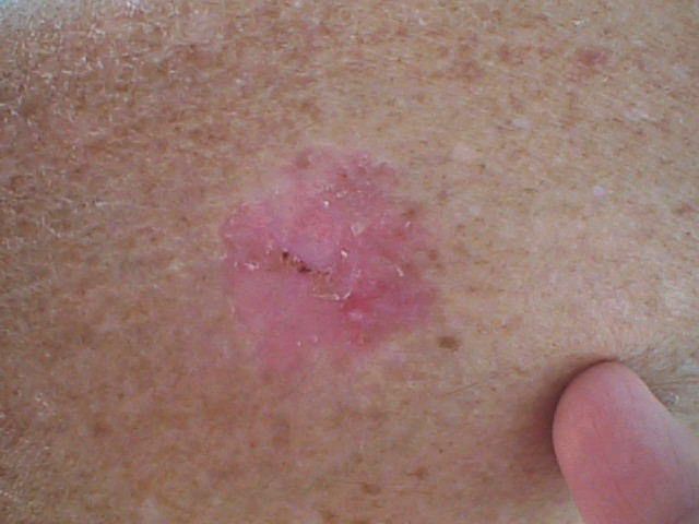
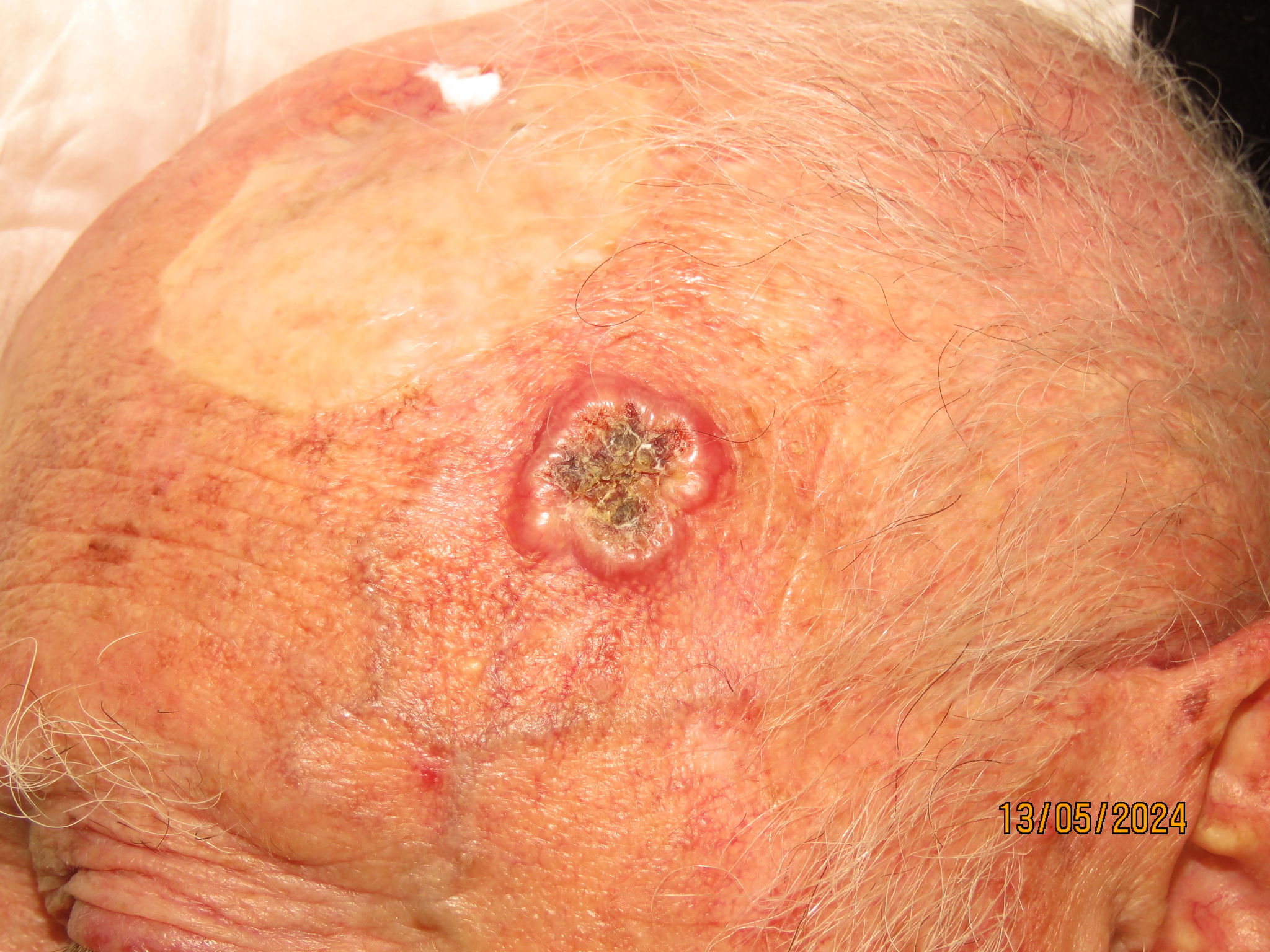
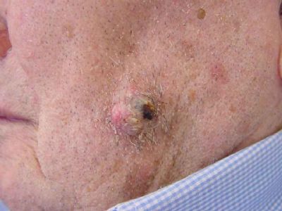
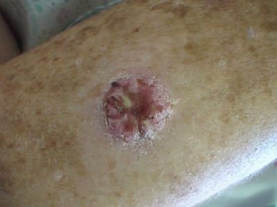
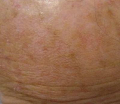
The most common form is the appearance of a bump/lump of tissue that grows on the surface of the skin quickly and is usually scarred in the center with stiff edges and a red halo surrounding it. This form is also the most malignant. Another form is a warty form covered with a layer of keratinous cortex and which usually does not heal. This form is considered less malignant but can change to the malignant form if not treated in time. Another common form is the one that appears in areas exposed to the sun as the development of precursor lesions called "actinic keratoses". These lesions, which can be detected as rough skin areas, sometimes with a slightly darker shade than the surrounding skin, and sometimes covered with a rough horny layer in areas exposed to the sun, often change to the form of warts. Another form is a reddish plaque, covered with thin scales, spreading horizontally across the skin, often with a sharp border from the surrounding healthy skin. This form is also called Bowen's tumor or Bowen's disease and SCC in situ, and is a very superficial form that never metastasizes but if not treated in time may change into a more invasive and violent form of the tumor.
In the pictures (from 1st to last): lump-shaped tumor/tissue lump, wart-shaped tumor, actinic keratoses, Bowen's type tumor

Where does the tumor usually appear?
The tumor can appear in any area of skin or mucous membranes, but the most common locations are on the skin of the face, especially on the lower lip and ears, as well as on the eyebrows, back of the hands, forearms and calves.
What are the dangers?
SCC is considered more dangerous than BCC due to the fact that it grows relatively quickly and due to the increased risk of metastases. The risk of metastases is related to the degree of malignancy of the tumor and such metastases often appear in early stages. The incidence rate of metastases to the lymph nodes is 4-5%. This type of cancer causes death in 1:100 patients. In addition to the risk of metastases, this is a tumor that can grow locally, bleed and destroy tissue, unless removed. When the tumor is located near vital structures such as the eyes, ears and nose, or near a nerve, the damage that may be caused can be severe unless the lesion was treated earlier and due to its tendency to progress relatively quickly it is advisable to treat it without delays.
Biopsy
Any skin lesion or wound that does not heal must be evaluated by a dermatologist who will examine and diagnose its nature, and in cases where the clinical diagnosis is not clear-cut will recommend a biopsy. A biopsy is a simple procedure that is usually performed in a clinic under local anesthesia, in which a small piece of tissue is sampled from the lesion for examination using the microscope. In some cases, the doctor will choose to remove the entire lesion in the first place, without waiting for the biopsy answer.
The treatment
After it has been diagnosed that it is a tumor of the SCC type, there are several treatment options:
Surgical excision - a small operation in which the lesion is removed in its entirety with a margin of skin that looks healthy about 0.5 cm wide. The edges of the wound are sewn together using a dermatosurgery technique. After the surgery, the removed tissue is sent to a pathologist who examines sample pieces of the tissue under the microscope and determines the nature of the tumor that was removed and whether the tumor was removed in its entirety.
Electrodesiccation and scraping - the tumor is scraped and removed using a "curette" (a sharp spoon-like device) and the base is treated with a burn with an electric needle (an unacceptable method for suspected SCC).
Cryosurgery - deep and thorough freezing of the tumor and its surroundings by direct spraying of liquid nitrogen (suitable only for the superficial forms of the SCC in situ or Bowen's tumor).
Local treatment - application of medicines such as "Efudix" ointment containing a therapeutic substance called 5-fluorouracil or "Aldara" cream containing a substance that activates the patient's immune system to treat the tumor. This treatment is particularly suitable for the superficial forms and "pre-cancerous" lesions.
Surgery using the MOHS method - surgery performed by a dermatologist who specializes in the method and is intended for special situations. The tumor is removed under local anesthesia and the tissue is examined under a microscope by the surgeon himself, to make sure that all the involved tissue has been removed. Additional layers of tissue are removed in stages until complete removal of the cancerous tissue is achieved. This method yields an extremely high success rate in removing the tumor but is not necessary for all types of tumors. The situations in which it is recommended to use this method - recurring tumors, partially removed tumors, large tumors or tumors in areas of special importance such as eyes, ears, nose and mouth. After the tumor has been completely removed, the wound is closed with aesthetic reconstruction methods or bandaged and left for gradual secondary healing.
Radiotherapy - Irradiation of the skin by electron beams or Alfa radiation with an intensity and spread adjusted to damage the tumor cells in the skin. According to experts, this treatment achieves a very respectable success rate, although there are no controlled studies to prove this data. The intensity of the treatment is adjusted to the location of the tumor, the surrounding tissues and the patient's tolerance - and possible to divide the total radiation dose into parts (fractions) that are given separately day after day, in a way that reduces the damage to healthy tissue and thus reduces the suffering for the patient. For protection during the treatment, a special structure is prepared from lead that blocks the radiation from reaching the skin that is not suspected to be involved. The treatment is particularly suitable for patients who are at high surgical risk, patients with several tumors (radiotherapy can treat several tumors at the same time and in relatively large areas), patients who fear surgery (painful needles, etc.) and special locations of tumors where surgery may cause severe damage that is difficult to repair, or one that would require a complex repair. At the same time, the need to go to the hospital numerous times for treatment is a limitation that causes quite a few patients who have difficulty with mobility to opt out of the option. The side effects of radiation are divided into immediate effects manifested by burns on the patient's skin, a wound where the tumor was, drying of adjacent mucous membranes (for example, the nasal mucosa when the irradiated tumor is located on the back of the nose) and late effects - relatively sensitive skin, which recovers slowly from bruises or surgeries, and there is also concern about the development of malignancies secondary to the radiation itself - a phenomenon that is considered particularly late and and may appear after a period of 15-20 years from the radiotherapy treatment. The treatment is included in the basket of treatments according to the health law.
Photodynamic therapy - this is a relatively new treatment in the basket of treatments. The treatment is intended for areas with severe sun damage and only superficial tumors of the BCC & SCC type and is given by applying a photosensitizer that is preferentially absorbed by abnormal skin cells such as tumor cells or cells damaged by sunlight. After about half an hour of contact with the substance, the skin sensitized by the substance (usually Amino Levulinic Acid) is exposed to visible light in the red range and the substance turns into oxygen radicals that destroy the cells in which it is ingested. This is how a specific injury to cancerous or pre-cancerous cells inside the skin is actually achieved. After the treatment, the condition is similar to a sunburn and within a few days the damaged cells die and peel off the skin, leaving the skin mostly uniform and free of cancerous and pre-cancerous lesions. This treatment is particularly suitable for the superficial forms and "pre-cancerous" lesions. Some argue that this treatment, when it fails to completely eliminate an SCC type tumor, turns it into a more active and aggressive tumor. This claim is also not supported by significant research evidence to date.
Chemotherapy - Systemic (systemic) chemotherapy treatment is indicated mainly for invasive and metastatic SCC tumors. This is a treatment given in an oncological setting and is accompanied by severe side effects and is usually also accompanied by radiation therapy for deep metastases of the tumor.
Will there be a scar? Since most tumors appear on the skin of the face, many patients are rightly concerned about the quality of the cosmetic result. When the tumors are small, conservative treatment methods yield excellent cosmetic results. In cases where the tumor requires excision surgery using the MOHS method, there are various aesthetic reconstruction options that in most cases yield an excellent cosmetic result. Even when the immediate result of the surgery is not optimal - it is often possible to use local means to improve and correct the result.
Follow up The studies show that when a person develops an SCC tumor they have a significantly increased risk of developing another skin cancer within 5 years. It is therefore important to persevere and meet follow-up and review appointments with the dermatologist, in order to detect any development as early as possible. The patient must be alert and aware of any non-healing wound that develops on the skin. It is important to regularly use sunscreens and be careful about sun exposure.
Associated risks Patients who have suffered from multiple SCC type tumors or other skin cancers such as basal cell carcinoma are at increased risk of melanoma. An SCC tumor cannot turn into a melanoma.
Prevention Since SCC tumors are often caused by UV radiation from the sun, avoiding sun exposure and protection from the sun's rays can help prevent it. Sun protection is especially important in children. Patients should perform a self-examination at regular intervals and undergo a general skin examination for the early detection of suspicious lesions at least once a year. It is important to seek shade, especially during the hours when the sun's radiation is especially strong (between 10 am and 4 pm). It is recommended to wear light, densely woven clothes, with long sleeves and pants, a wide-brimmed hat and sunglasses. It is recommended to apply broad-spectrum sunscreen cream that protects against both UVA and UVB radiation with a protection factor of at least 15 SPF. Sunscreen must be reapplied every two hours in the sun and even on cloudy days.
Dr. Gilead carries out an orderly follow-up of patients at increased risk of skin cancer of all types in his clinic, using dermoscopy.
Preventive treatments are performed in patients who are at risk and in patients who develop new tumors -
Dr. Gilead removes the tumors using the MOHS method or regular surgery or refers the patients to another appropriate treatment.


